The most basic and functional unit of life is called a cell. These cells
make up every organism on earth. First discovered by
Robert Hooke
when examined some
cork
, these were named
cells because their structure looked like
honeycomb cells.
These cells are not functional on their own but need several organelles to do
the heavy lifting. Each organelle has different functions but these come
together to serve the common purpose of the cell.
I am going to document these organelles in detail, from their structure,
functions to their correspondence with other cell organelles. But first we need
to look at cell theory, which is the basis of modern biology.
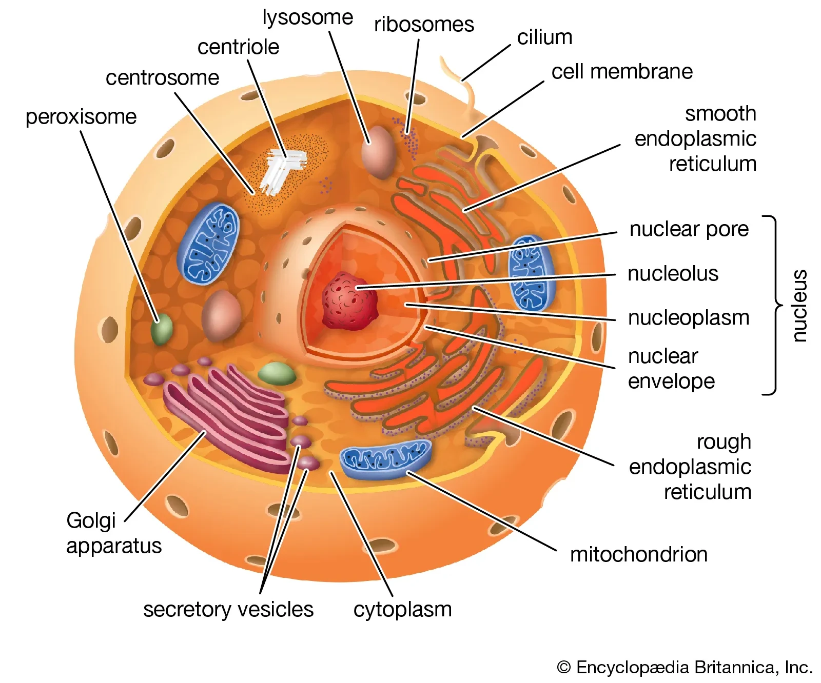
An Animal Cell. Courtesy : Encyclopædia Britannica
Cell Theory
Two German scientists, Matthias Schleiden and Theodor Schwann worked together and made discoveries about cells and their role in all organisms.
Matthias Schleiden first proposed in 1839 that every plant is made up cells,
while Schwann extended this statement to animals in the same year. However, they
both disagreed on how cells come to be. Schleiden believed that cells came from
free cell formation, i.e., they crystallized into existence out of nowhere.
Schleiden’s claim was refuted by
Rudolf Virchow
who gave the idea
that cells came from pre-existing cells. Virchow is believed to have
plagiarized
the work of
a polish scientist Robert Remak
.
These gave us the three principles of cell theory.
Principles of Cell Theory
- All organisms are composed of one or more cells.
- The cell is the most basic unit of structure and organisation in organisms.
- Cells come from pre-existing.
Classification of Cells
Cells, on the basis of their nuclear material, are divided into two types :
1. Prokaryotic Cells - Cells which don’t possess a well-defined nucleus and
organelles are called prokaryotes. Here pro means primitive/before in Greek
and karyon means kernel, sopro + karyon = prokaryote (primitive cell).
Prokaryotic cells are generally cells of bacteria, viruses, fungi or other such single-celled organisms like amoeba.
2.Eukaryotic Cells - Cells which have a well-defined, membrane-bound nucleus
and membrane-bound organelles are called eukaryotes. Here eu means well/good
in Greek and—as previously mentioned—karyon means kernel, so
eu + karyon = eukaryote ( Well defined cell).
Eukaryotic cells are generally cells of complex, multi-cellular organisms like monkeys.

Diagram of a Prokaryotic Cell
Difference in Prokaryotes and Eukaryotes
Sourced from wikipedia.
| Prokaryotes | Eukaryotes | |
|---|---|---|
| Size | ~1-5 μm | ~10-100 μm |
| Nucleus | Nucleoid, no true nucleus | contains true, double membrane-bound nucleus |
| Organism | Typically single celled | single as well as multi-cellular |
| Cell Division | Binary Fission | Mitosis / Meiosis |
| Organelles | No specialized, membrane-bound organelles | specialized, membrane-bound organelles are present |
Classification of Eukaryotic Cells
Eukaryotic cells are further divided according to the type of organism they belong to. These are of two types:
Animal Cells
Plant Cells
Animal Cells and Plant Cells
Eukaryotic cells belonging to members of the animal kingdom are called
animal cells.
Similarly, cells belonging to members of the plant kingdom are called
plant cells.
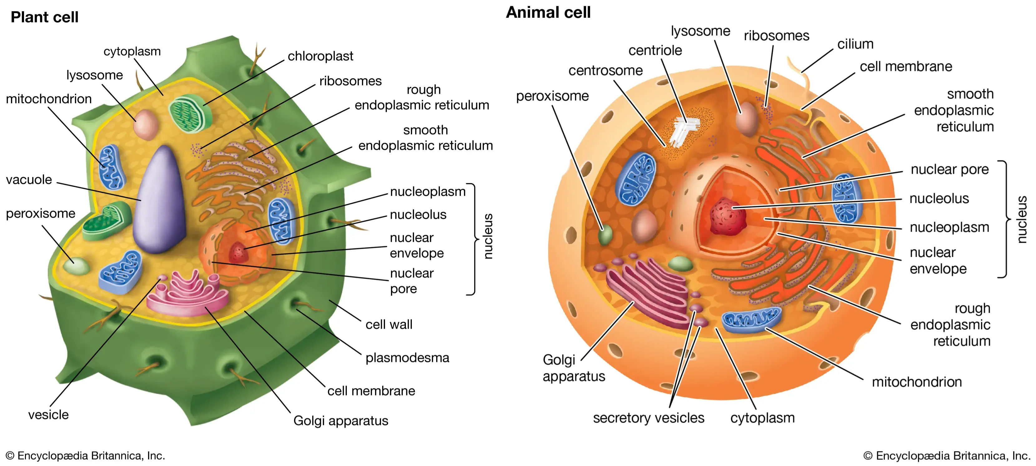
Comparison of Plant and Animal Cells. Courtesy : Encyclopædia Britannica
This diagram shows both an animal cell and a plant cell side by side. It may be observed that both have most of the organelles in common (some with a little difference) and a few unique ones. These organelles are what power these cells.
Cell Components
I will categorize others as “components” because they don’t quite fit the category of organelles. These are :
- Cell Membrane
- Cell Wall
- Cytoplasm
Cell Membrane
The outermost-in animal cells and the innermost in plant cells- layer is called
the cell membrane. It’s a selectively permeable membrane, as in it allows some
substances such as
H2O
,
CO2
etc., pass while blocking
undesirable objects like hostile organisms.
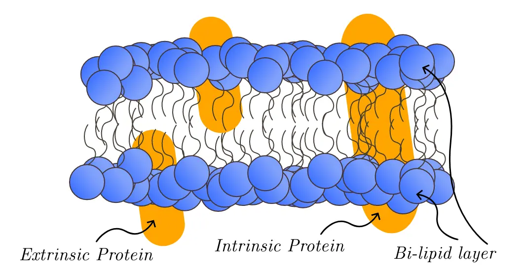
Cell Membrane Layers
Structure of the Cell Membrane
The cell membrane is made up of two layers of phospholipids, also called a
bi-lipid layer, which have proteins between them.
The proteins which fully penetrate the bi-lipid layer are called intrinsic
proteins, while the ones which only fully penetrate one of the two layers are
called extrinsic proteins.
Functions of the Cell Membrane
- Intrinsic proteins help in the exchange of substances to and from the cell.
- Extrinsic proteins while not fully penetrating the bi-lipid layer, it also helps in transport of materials in times of distress. It also alerts the cell of the dangers outside like viruses, hostile bacteria etc.
Cell Wall
The outermost boundary of plant, algae and fungi cells is called a cell wall.
- It’s a dead component.
- Has high tensile strength.
- It’s freely permeable.
Plant cells are composed of cellulose, hemicellulose and pectin.
Cytoplasm
The area inside the cell is filled with a viscous, gelatinous fluid which is called cytoplasm. It helps in the transport of cellular substances like proteins, lysosomes etc. inside the cell.
Composition of Cytoplasm
| Substance | % |
|---|---|
| Oxygen | 62 |
| Carbon | 20 |
| Hydrogen | 10 |
| Nitrogen | 3 |
| Calcium | 2.5 |
| Other essential materials | 2.5 |
Parts of Cytoplasm
Cytosol - The translucent fluid in which cell organelles are suspended, it make up 70% of the cytoplasm.
Cytoskeletal - The fibrous strands which give the cell a proper shape, adjust the cell organelles position and aid in the transport of vesicles. These are only found in eukaryotes.
Functions of Cytoplasm
To give the cell a proper shape via the cytoskeletal.
To help in dissolving in water-soluble substances.
It aids in the transfer of cellular material inside the cell.
Cell Organelles
Cell organelles are sub-units of a cell which generally have a specialized function. There are quite a few of these, each with a different structure, size, function etc. The organelles covered here are going to be:
Common organelles
Organelles specific to plant cells
Nucleus
The nucleus is a spherical, double membrane-bound organelle located at the periphery in plant cells and at the center in animal cells, it’s the most important cell organelle in the cell as it controls all cellular functions.
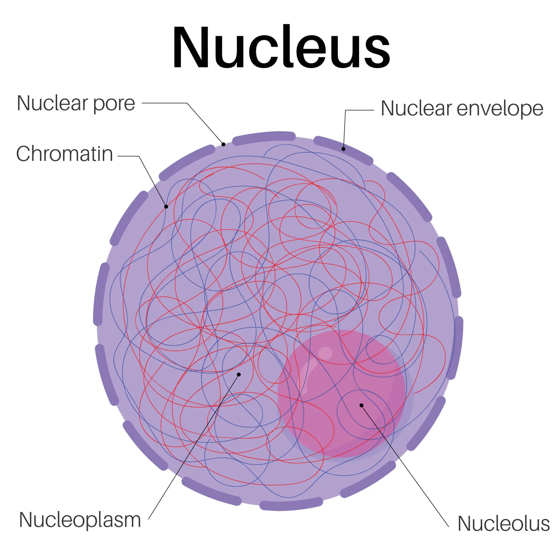
A Nucleus Diagram
- The nucleus is what differentiates eukaryotes and prokaryotes.
- It controls the metabolic activities of the cell.
Nuclear Membrane
This is a double layered membrane which separates the nucleus from the cytoplasm of the cell. This protects the cell from foreign objects and organisms.
Nuclear Envelopes & Pores
Nucleus pores are small openings in the nuclear membrane, these allow the entry and exit of macromolecules like ribosome. Envelopes are then, the pieces of the dashed membrane.
Chromatin
Long threads made up of DNA and proteins carrying genetic data necessary for cell division. These condense into gene-carrying chromosomes during cell division.
Nucleolus
The spherical structure present at the center of the nucleus. It’s not membrane bound and is responsible for the synthesis of ribosome.
Endoplasmic Reticulum
The Endoplasmic Reticulum is a contains membrane organelle made up of flattened sacs, covering the space around the nucleus.
It’s found in most eukaryotic cells.
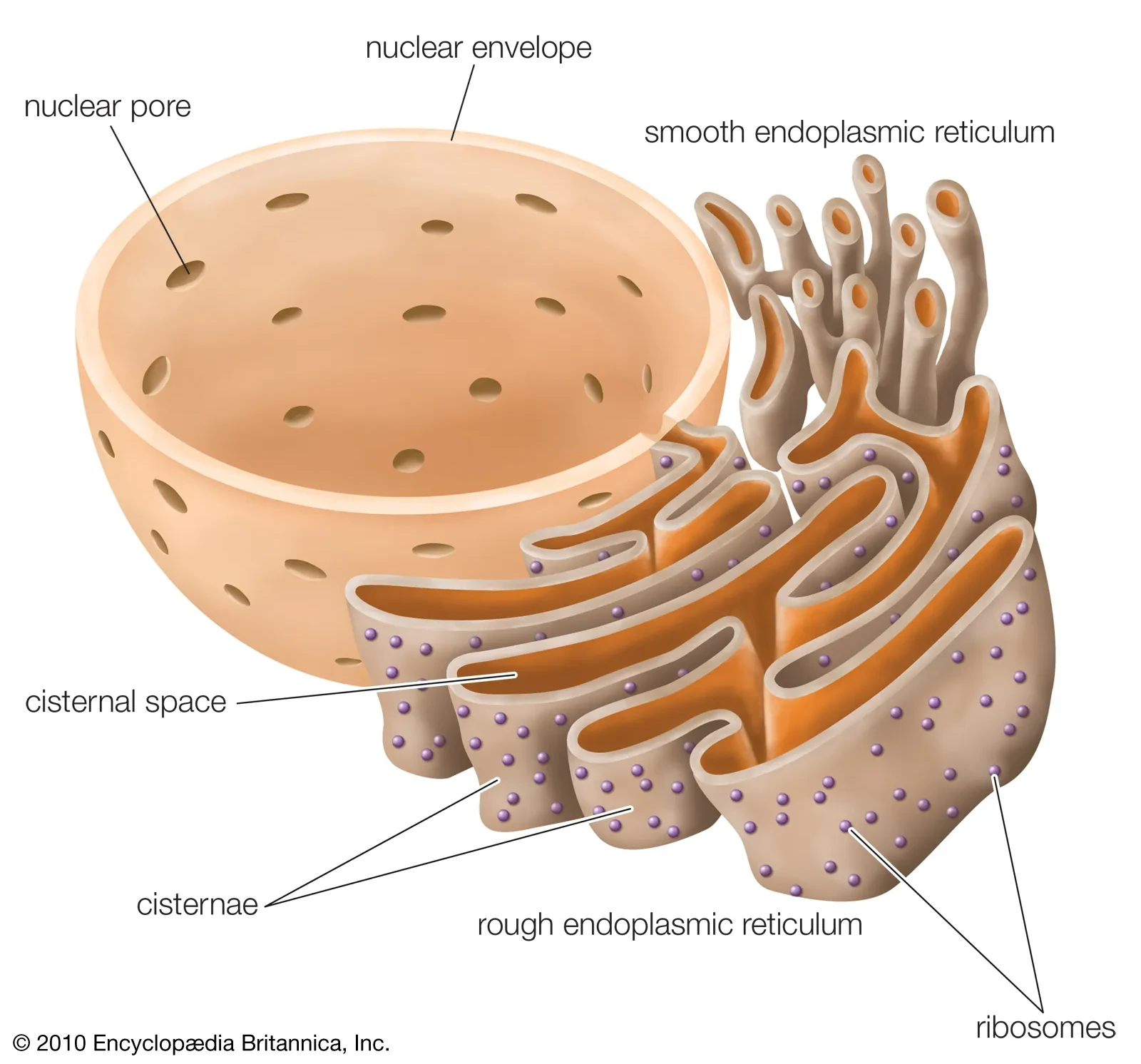
Endoplasmic Reticulum. Courtesy : Encyclopædia Britannica
According to appearance, the E.R. is divided into two types :
- Rough Endoplasmic Reticulum
Named so due to appearance caused by embedded ribosomes which enable it to make protein.
- Smooth Endoplasmic Reticulum
Not having embedded ribosomes gives this part of the E.R. a smooth appearance,
due to the lack of ribosomes, it manufactures lipids.
Functions of the Endoplasmic Reticulum
Under Rough Endoplasmic Reticulum
- Synthesis of protein with the help of the ribosomes.
- Modification of proteins.
- Transportation of proteins to different sites of the cell via packaging in vesicles.
Under Smooth Endoplasmic Reticulum
- Synthesis of lipids.
- Transportation of lipidids.
- Secretion of hormones via the endocrine glands.
- Detoxification and filtering of consumed substances, specially during alcohol and drug overdose.
The Golgi Apparatus
The Golgi Apparatus or the golgi apparatus is a membrane-bound cell organelle
made up of flat, stacked sacs called cistarnae. It’s found in most eukaryotic
cells.

Golgi Body. Courtesy : Encyclopædia Britannica
The Golgi Network
It has three components and two networks, namely:
- Components
Cis : The face closest to the E.R.
Medial : Middle part of the golgi apparatus, located at medium distance from the E.R.
Trans : The face closest to the E.R.
- Networks
Cis Network
Trans Network
The networks belong to their respective components and are composed of multiple cistarnae. Here’s a youtube video explaining the networks in detail.
Functions of the Golgi Apparatus
The proteins and lipids manufactured by the E.R. go into the golgi apparatus to be :
1. Modified : It modifies the proteins/lipids by adding other components such as other types of proteins and lipids.
2. Packaged & Transported : It packages its modified proteins/lipids into vesicles and transports them across the cell.
Since the vesicles forms out of the golgi itself, its essentially always forming.
Mitochondria
As you most probably already know, mitochondria is the powerhouse of the cell.
It’s a vital organelle for eukaryotes (it’s only found in eukaryotic cells), it’s double membrane-bound, it uses oxygen (through aerobic respiration) to dissolve nutrients into energy that’s usable by the cell, into adenosine triphosphate or ATP.
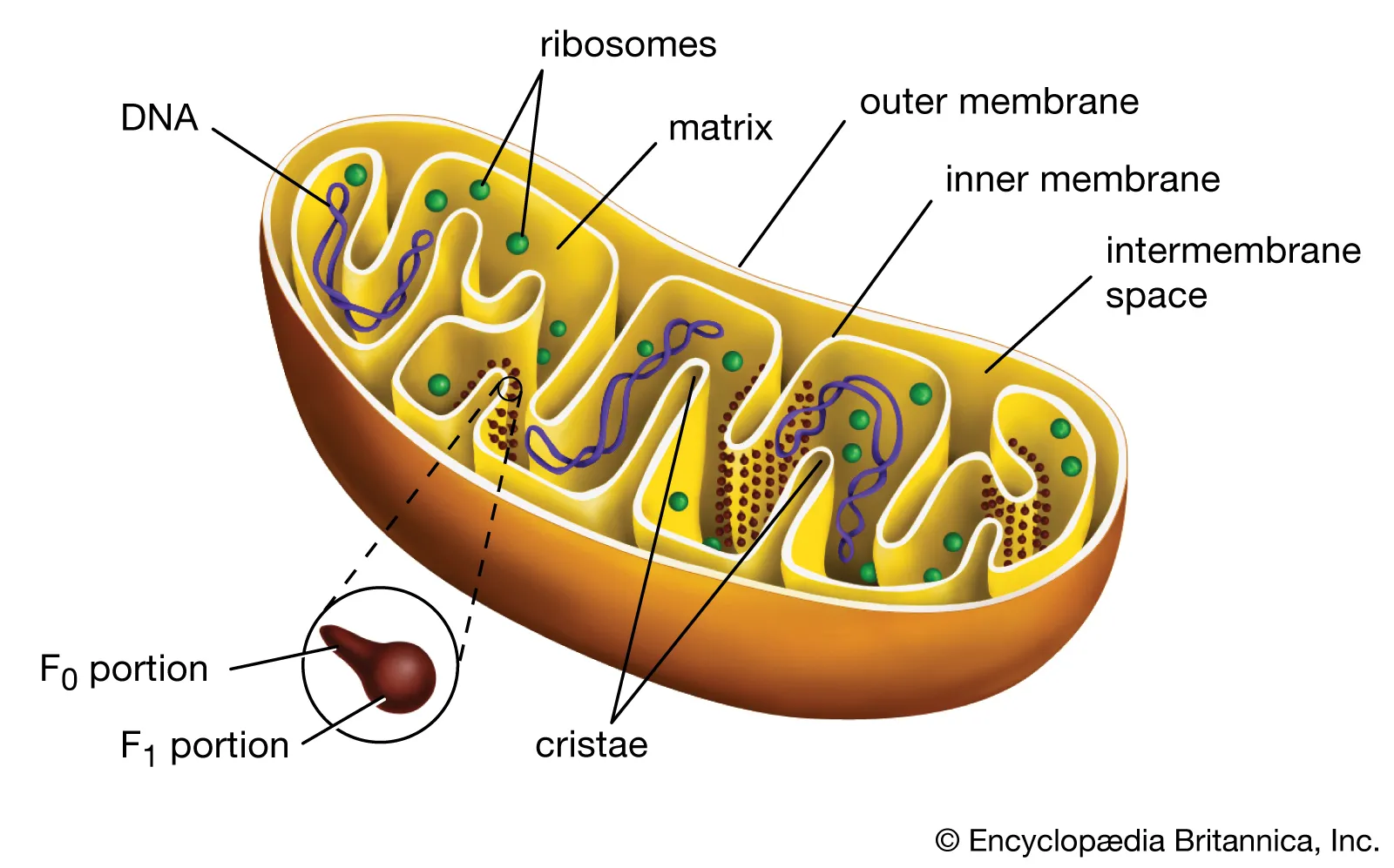
Mitochondria. Courtesy : Encyclopædia Britannica
Structure of the Mitochondria
It’s bean shaped with two membranes -
Inner Membrane
Outer Membrane
The inner membrane is folded towards the inside making a finger-like structure
called cristae. These folds host a particle which is essential to the
production of ATP. These are called F1 particles or oxysomes. That’s the
reason these finger-like folds exist, having these increases the surface area on
which the F1 particles are hosted, therefore increasing the ATP production.
It also houses a matrix in the inner membrane which is a liquid containing different types of enzymes, ribosomes and other proteins.
Lysosome
Lysosome is a membrane-bound organelle present in many eukaryotes. It carries various digestive enzymes inside its membrane for varied purposes. Its structure is quite simple, it’s just a membrane-bound spherical object filled with digestive enzymes.
Functions of the Lysosome
To digest foreign organisms which enter the cell.
To digest harmful foreign substances.
To release digestive enzymes into the cell if it becomes diseased and may harm surround cells, this is also why it’s called suicidal bag.
Vacuole
Vacuoles are some of the more noticeable category of organelles. They exist in both animal cells and plant cells. They’re generally formless and expand/contract as the cell needs it to. They are far bigger in plant cells than their animal cell counterparts, as can be seen here.
Functions of Vacuoles
They are mostly containers for different types of storage.
- Isolating materials that might be harmful or a threat to the cell.
- Containing waste products.
- Containing water in plant cells.
- Maintaining internal hydrostatic pressure or turgor within the cell during osmosis.
Plastids
Plastids are a group of double membrane-bound organelles found only in plant cells and algae. We’ll cover only 3 types of plastids out of the eight.
They are :
1. Chromoplast
2. Chromoplast
3. Leucoplast
Chromoplast
These kinds of plastids contain a non-green pigment (usually brownish to reddish), they have irregular shapes and they don’t change into other types of plastids.
- They give color to the non-green parts of a plant like the flowers, fruits, roots etc.
Chloroplast
Chloroplasts are perhaps the most important type of plastids. They contain
chlorophyll, which gives plants their distinctive green color and enables them
to manufacture food via photosynthesis.
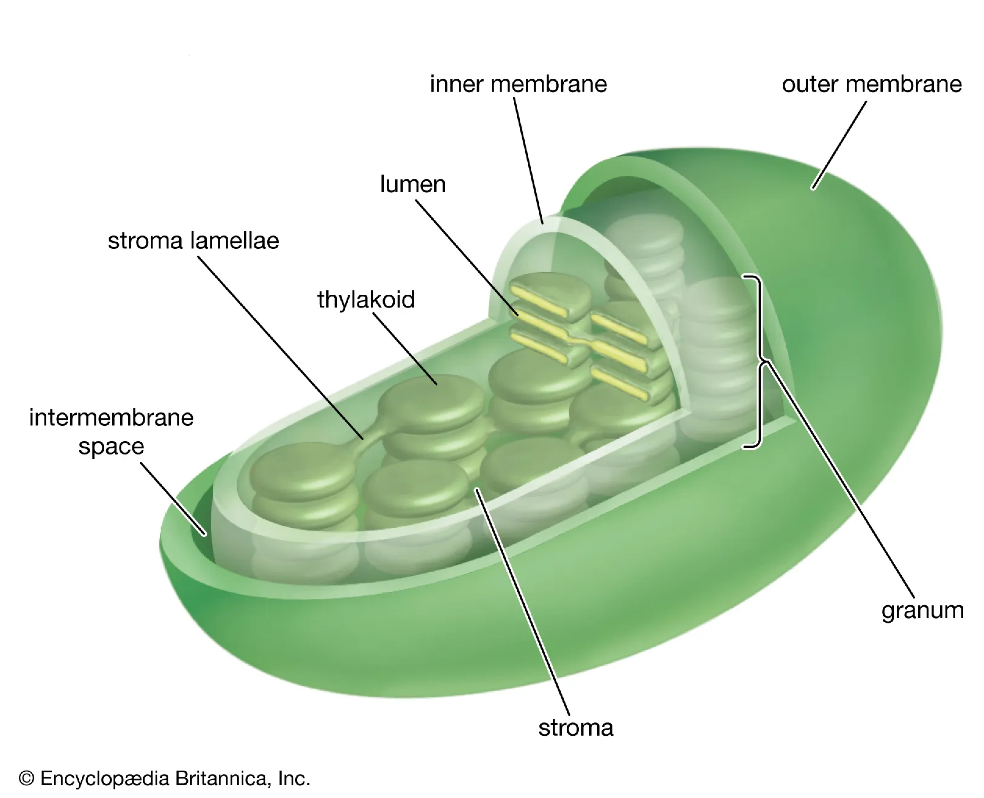
A Chloroplast Visualized in 3D. Courtesy : Encyclopædia Britannica
The above diagram shows a chloroplast with the following :
1. Thylakoid : These are membrane-bound compartments which are shaped like
sacs, (Thylakoid comes from Greek thylakos, which means sac) they are the
sites of photosynthesis.
2. Grana : A bunch thylakoids when stacked together are called grana (singular : granum).
3. Stroma : Is the homogeneous in which grana exist.
4. Lamella : Lamella simply connects different grana.
Leucoplast
These are colorless, that is, they contain no pigment. They are spherical in shape and store food like starch, lipids and protein.
Functions of Plastids
1. Chloroplast
- Perform photosynthesis to manufacture food.
- Give color to leaves, shoots, stem etc.
2. Chromoplast
- Give color to flowers, fruits, roots etc. to attract animals for pollination and/or seed dispersal.
3. Leucoplast
- To store starch, lipids and protein.
Until next time!